Pregnancy ultrasound scans help doctors monitor a baby’s development, estimate the due date, assess the pregnancy, and identify certain conditions that may require further evaluation. Knowing when each pregnancy ultrasound is usually performed can help expectant parents prepare for antenatal appointments and understand the purpose of each scan.
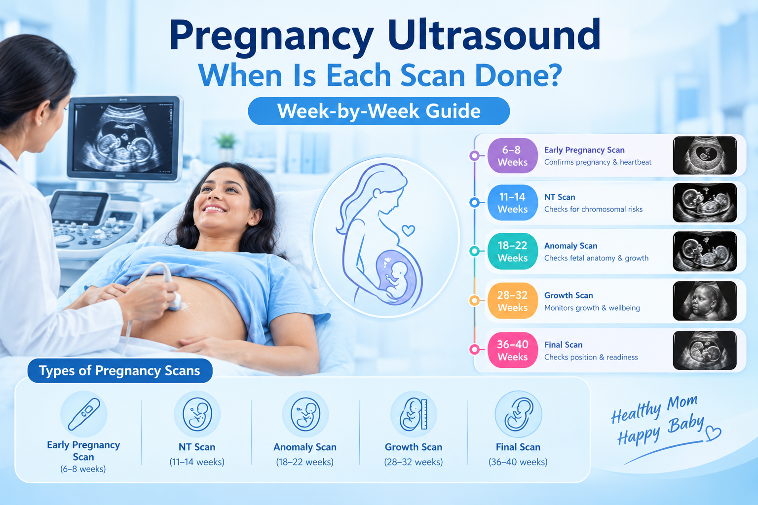
The pregnancy ultrasound schedule may include an early pregnancy or dating scan, a first-trimester nuchal translucency (NT) scan, a second-trimester anomaly scan, and additional growth or Doppler scans when medically indicated. The exact timing and number of scans depend on your pregnancy, medical history, and your doctor’s recommendations.
For expert guidance on pregnancy scans and antenatal care, consult the Best Gynecologist in Hassan to understand which ultrasound examinations are appropriate for your pregnancy.
What Is a Pregnancy Ultrasound?
A pregnancy ultrasound is a medical imaging test that uses sound waves to produce images of the developing baby, uterus, and surrounding structures. It does not use ionising radiation.
Ultrasound scans can help confirm the location of a pregnancy, estimate gestational age, assess fetal growth, identify multiple pregnancies, examine the baby’s anatomy, and evaluate the placenta and amniotic fluid.
Some scans are routinely offered as part of antenatal care, while others are recommended when a doctor needs more information about a particular concern.
For an overview of prenatal care, read Karna Hospital’s guide to pregnancy care month by month.
Pregnancy Ultrasound Schedule by Trimester
The timing of pregnancy ultrasound scans can vary depending on local clinical protocols and individual needs. The following schedule explains common scans and their purposes.
| Pregnancy stage | Common scan | Main purpose |
| Early pregnancy | Dating or viability scan, when indicated | Assess pregnancy location, development and gestational age |
| 11–14 weeks | NT scan and first-trimester screening, when chosen or recommended | Assess early development and screen for certain chromosomal conditions |
| 18–22 weeks | Anomaly or fetal anatomy scan | Examine the baby’s developing organs and physical structures |
| Third trimester | Growth scan, when indicated | Assess fetal growth, position, fluid and other clinical factors |
| Any trimester, when needed | Additional ultrasound or Doppler | Investigate specific concerns and guide care |
Not every pregnancy requires every scan listed here. Your obstetrician will recommend a schedule based on your circumstances.
1. Early Pregnancy Ultrasound: When Is the First Scan Done?
An early pregnancy ultrasound may be recommended during the first trimester, depending on your menstrual history, symptoms, and clinical needs. In some cases, a scan is performed at around 6–9 weeks, although timing varies.
What does the first pregnancy ultrasound check?
An early pregnancy scan may help:
- Confirm that the pregnancy is located inside the uterus.
- Assess early pregnancy development and cardiac activity when visible.
- Estimate how many weeks pregnant you are.
- Help calculate or confirm the expected due date.
- Check whether you are carrying one baby or more than one.
- Investigate pain or bleeding when clinically indicated.
If the scan is performed very early, it may not yet show all the expected structures. Your doctor may recommend a repeat scan rather than drawing conclusions from an early examination alone.
For practical guidance during the early weeks, see Karna Hospital’s first trimester pregnancy care guide.
2. NT Scan: When Is the 12-Week Pregnancy Scan Done?
The nuchal translucency (NT) scan is generally performed between 11 weeks and 13 weeks plus 6 days, according to the relevant screening protocol. It measures the fluid space at the back of the baby’s neck.
The scan may form part of first-trimester screening for certain chromosomal conditions, including Down syndrome. Depending on the screening programme, it may be combined with blood tests and other assessments.
What does the NT scan check?
The scan may assess:
- The baby’s size and development at this stage.
- Gestational age.
- Nuchal translucency measurement.
- Certain early structural features.
- Whether further screening or assessment should be discussed.
An NT scan is a screening test, not a definitive diagnosis. A result indicating a higher chance of a condition does not necessarily mean the baby has that condition. Your clinician can explain the results and discuss any recommended follow-up tests.
3. Anomaly Scan: When Is the 20-Week Pregnancy Scan Done?
The anomaly scan, also called the fetal anatomy scan or TIFFA scan in many Indian clinical settings, is generally performed between 18 and 22 weeks of pregnancy.
It is one of the most detailed routine ultrasound examinations because it evaluates the baby’s developing anatomy.
What does the anomaly scan check?
The sonographer examines visible structures, which may include:
- The baby’s brain and head.
- The face and spine.
- The heart and chest.
- The stomach, kidneys and bladder.
- The arms, legs, hands and feet.
- The placenta and amniotic fluid.
- Measurements used to assess fetal size.
The scan can identify some structural abnormalities, but it cannot detect every possible condition. Sometimes the baby’s position or other technical factors make it difficult to obtain all the required views, and a repeat scan may be recommended.
The anomaly scan is not the same as the NT scan. The NT scan is part of first-trimester screening, whereas the anomaly scan focuses on a more detailed examination of fetal anatomy in the second trimester.
4. Third-Trimester Ultrasound: When Is a Growth Scan Done?
A third-trimester growth scan may be recommended when the doctor needs to assess the baby’s growth or investigate a particular clinical concern. There is no single week that applies to every pregnancy.
Depending on the reason for the examination, a growth scan may assess:
- The baby’s estimated size and growth pattern.
- The baby’s position in the uterus.
- The amount of amniotic fluid.
- The location of the placenta when relevant.
- Other findings that may affect pregnancy management.
A growth scan can be particularly useful when the doctor suspects that the baby is smaller or larger than expected, or when maternal health conditions require closer monitoring.
However, a third-trimester growth scan is not automatically required at the same week for every uncomplicated pregnancy. Your obstetrician will determine whether and when it is appropriate.
5. Doppler Scan During Pregnancy: When Is It Needed?
A Doppler ultrasound assesses blood flow through selected blood vessels. During pregnancy, it may be recommended when the doctor needs additional information about circulation between the placenta and baby or other aspects of fetal well-being.
A Doppler scan may be used in selected situations, including suspected fetal growth restriction or certain high-risk pregnancies.
It is not necessarily part of every routine pregnancy ultrasound schedule. The timing and type of Doppler examination depend on the clinical reason for testing.
How Many Ultrasound Scans Are Needed During Pregnancy?
There is no universal number of ultrasound scans required for every pregnancy.
Some antenatal programmes routinely offer a dating scan and a second-trimester anomaly scan, while other clinical pathways may include additional examinations based on local guidelines and individual needs.
You may need more scans if your doctor needs to monitor fetal growth, investigate bleeding or pain, assess the placenta, or manage a pregnancy-related medical condition.
The purpose of ultrasound is to provide useful clinical information—not simply to reach a particular number of scans. Follow the schedule recommended by your treating clinician.
For pregnancy monitoring and comprehensive medical support, you can explore the services offered by the Best multispeciality hospital in Hassan.
Is Pregnancy Ultrasound Safe for the Baby?
Diagnostic ultrasound uses sound waves rather than ionising radiation and has a long history of use in obstetric care. When performed by trained professionals for appropriate medical reasons, it is considered a standard part of pregnancy assessment.
Scans should be performed by qualified healthcare professionals using appropriate equipment and protocols. Ultrasound examinations should be medically justified, and unnecessary or prolonged scanning solely for keepsake images should be avoided.
If you have concerns about the safety, timing, or need for a particular scan, discuss them with your obstetrician.
How Can You Prepare for a Pregnancy Ultrasound?
Preparation depends on the type and timing of the scan.
Before your appointment:
- Confirm whether you need a full bladder for the examination.
- Carry previous ultrasound reports and relevant medical records.
- Follow the instructions provided by the hospital or imaging centre.
- Ask your doctor whether the scan includes any screening tests that require separate consent.
- Discuss any questions about the purpose or limitations of the scan.
Some early pregnancy scans may require a full bladder, while other scans may not. Follow the specific instructions given for your appointment.
Frequently Asked Questions About Pregnancy Ultrasound
When is the first ultrasound done in pregnancy?
An early pregnancy scan may be performed around 6–9 weeks when indicated, but timing varies. The scan may help assess pregnancy location, early development, and gestational age.
When is the 12-week scan done?
The NT scan is generally performed between 11 weeks and 13 weeks plus 6 days, depending on the screening protocol.
When is the 20-week anomaly scan done?
The fetal anatomy or anomaly scan is usually performed between 18 and 22 weeks of pregnancy.
How many ultrasound scans are needed during pregnancy?
The number depends on local antenatal protocols and individual clinical needs. Additional scans may be recommended when closer monitoring is necessary.
Is a growth scan necessary in the third trimester?
Not necessarily. A growth scan may be recommended when your doctor needs to assess fetal growth, amniotic fluid, or other concerns. The decision depends on your pregnancy and medical history.
Can an ultrasound detect all birth defects?
No. Ultrasound can identify many structural abnormalities, but not every condition is detectable. Some findings may also require additional tests or specialist assessment.
Conclusion
Understanding the pregnancy ultrasound schedule can help you prepare for each stage of antenatal care. An early scan may assess pregnancy location and dating, an NT scan may contribute to first-trimester screening, and an anomaly scan examines the baby’s developing anatomy. Growth and Doppler scans may be recommended when additional monitoring is clinically appropriate.
The right schedule depends on your pregnancy and your doctor’s assessment. Attend your recommended antenatal appointments, keep your scan reports, and discuss any concerns with your healthcare professional.
For guidance on pregnancy monitoring and maternal care, explore Karna Hospital’s pregnancy care month-by-month guide and consult your obstetrician about the scans appropriate for your pregnancy.